Chemoselective reaction of methoxyaminomethyl BODIPYs with unprotected carbohydrates: a powerful tool for accessing BODIPY neoglycosides

The neoglycosylation of methoxyaminomethyl-appended BODIPYs with unprotected reducing mono-, di-, and trisaccharides produces, in a regio- and stereoselective manner, cyclic N-glycosyl-N-methoxy–BODIPY conjugates, as a relevant class of neoglycosides that display excellent photophysical characteristics in pure water, even at high dye concentrations. In addition, the cellular uptake of some of the neoglycosylated BODIPYs has been confirmed via fluorescence microscopy, and a BODIPY–acarbose conjugate showed comparable enzymatic inhibitory activity to acarbose for two different α-amylases: A. oryzae α-amylase (AOA) and human salivary α-amylase (HSA).

DOI
https://doi.org/10.1039/D4QO00886C

Europium–tannic acid nanocomplexes devised for bone regeneration under oxidative or inflammatory environments

Europium ions (Eu3+) are gaining attention in the field of regenerative medicine due to increasing evidence of their osteogenic properties. However, inflammatory and oxidative environments present in many bone diseases, such as osteoporosis or rheumatoid arthritis, are known to hinder this regenerative process. Herein, we describe a straightforward synthetic procedure to prepare Eu3+–tannic acid nanocomplexes (EuTA NCs) with modulable physicochemical characteristics, as well as antioxidant, anti-inflammatory, and osteogenic properties. EuTA NCs were rationally synthesized to present different contents of Eu3+ on their structure to evaluate the effect of the cation on the biological properties of the formulations. In all the cases, EuTA NCs were stable in distilled water at physiological pH, had a highly negative surface charge (ζ ≈ −25.4 mV), and controllable size (80 < Dh < 160 nm). In vitro antioxidant tests revealed that Eu3+ complexation did not significantly alter the total radical scavenging activity (RSA) of TA but enhanced its ability to scavenge H2O2 and ferrous ions, thus improving its overall antioxidant potential. At the cellular level, EuTA NCs reduced the instantaneous toxicity of high concentrations of free TA, resulting in better antioxidant (13.3% increase of RSA vs. TA) and anti-inflammatory responses (17.6% reduction of nitric oxide production vs. TA) on cultures of H2O2– and LPS-stimulated macrophages, respectively. Furthermore, the short-term treatment of osteoblasts with EuTA NCs was found to increase their alkaline phosphatase activity and their matrix mineralization capacity. Overall, this simple and tunable platform is a potential candidate to promote bone growth in complex environments by simultaneously targeting multiple pathophysiological mechanisms of disease.

DOI
https://doi.org/10.1039/D4TB00697F

Ti/Ta-based composite polysaccharide scaffolds for guided bone regeneration in total hip arthroplasty

Guided bone regeneration can play an important role in orthopedic applications. This work presents the synthesis and characterization of composite scaffolds based on polysaccharides loaded with microparticles of titanium or tantalum as novel materials proposed for composite systems with promising characteristics for guided bone regeneration. Ti/Ta composite scaffolds were synthesized using chitosan and gellan gum as organic substrates and crosslinked with oxidized dextran resulting in stable inorganic-organic composites. Physico-chemical characterization revealed a uniform distribution of metal nanoparticles within the scaffolds that showed a release of metals lower than 5 %. In vitro biological assays demonstrated that Ta composites exhibit a 2 times higher ALP activity than Ti and a higher capacity to support the full differentiation of human mesenchymal stem cells into osteoblasts. These results highlight their potential for bone regeneration applications.

1.
García-Robledo, H. et al. Ti/Ta-based composite polysaccharide scaffolds for guided bone regeneration in total hip arthroplasty. International Journal of Biological Macromolecules 271, 132573 (2024). Cite

Nanofibrous ε-Polycaprolactone Matrices Containing Nano-Hydroxyapatite and Humulus lupulus L. Extract: Physicochemical and Biological Characterization for Oral Applications

Oral bone defects occur as a result of trauma, cancer, infections, periodontal diseases, and caries. Autogenic and allogenic grafts are the gold standard used to treat and regenerate damaged or defective bone segments. However, these materials do not possess the antimicrobial properties necessary to inhibit the invasion of the numerous deleterious pathogens present in the oral microbiota. In the present study, poly(ε-caprolactone) (PCL), nano-hydroxyapatite (nHAp), and a commercial extract of Humulus lupulus L. (hops) were electrospun into polymeric matrices to assess their potential for drug delivery and bone regeneration. The fabricated matrices were analyzed using scanning electron microscopy (SEM), tensile analysis, thermogravimetric analysis (TGA), FTIR assay, and in vitro hydrolytic degradation. The antimicrobial properties were evaluated against the oral pathogens Streptococcus mutans, Porphyromonas gingivalis, and Aggregatibacter actinomycetemcomitans. The cytocompatibility was proved using the MTT assay. SEM analysis established the nanostructured matrices present in the three-dimensional interconnected network. The present research provides new information about the interaction of natural compounds with ceramic and polymeric biomaterials. The hop extract and other natural or synthetic medicinal agents can be effectively loaded into PCL fibers and have the potential to be used in oral applications.

Nanocarrier of α-Tocopheryl Succinate Based on a Copolymer Derivative of (4,7-dichloroquinolin-2-yl)methanol and Its Cytotoxicity against a Breast Cancer Cell Line

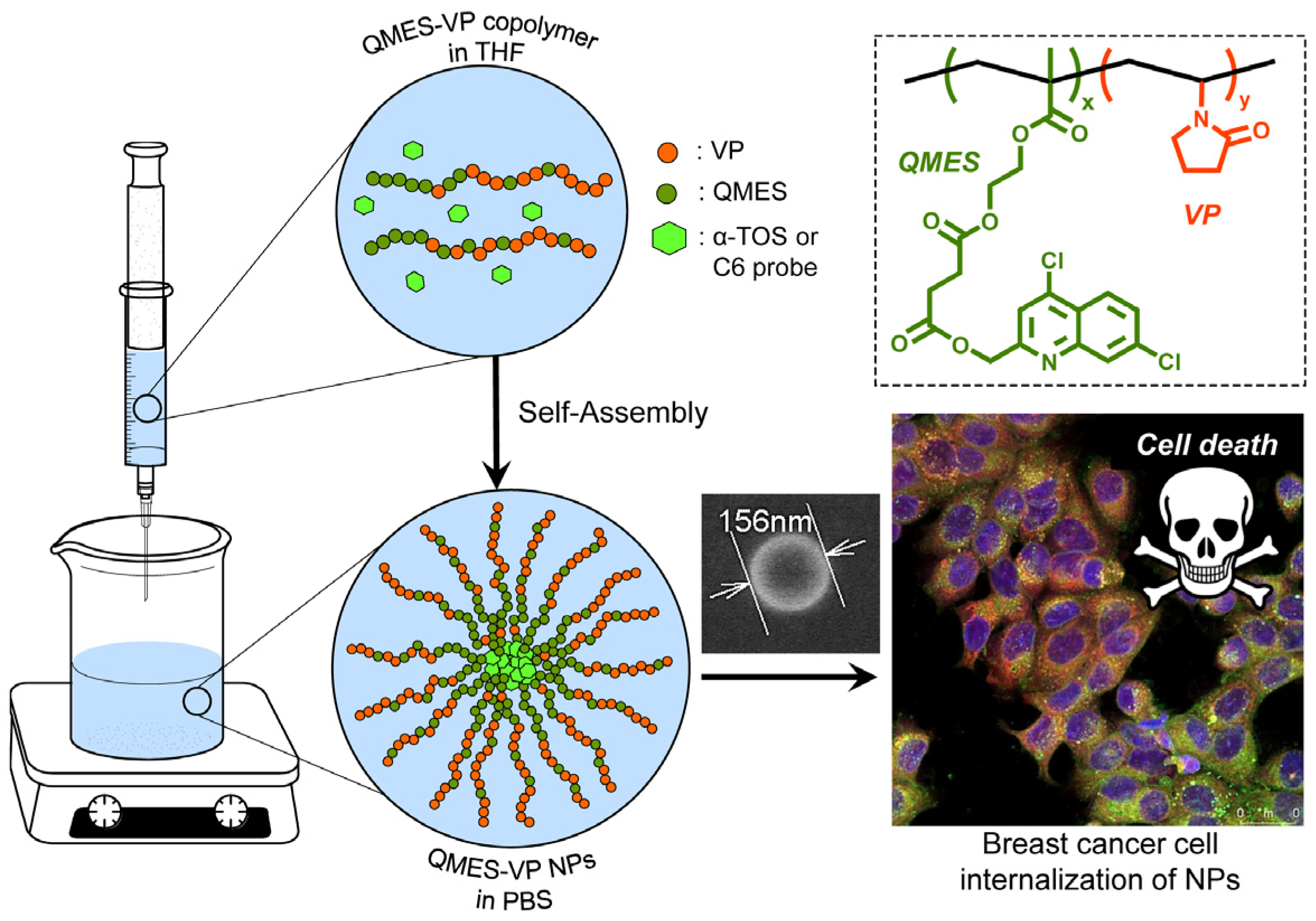
In order to improve the water solubility and, therefore, bioavailability and therapeutic activity of anticancer hydrophobic drug α-tocopherol succinate (α-TOS), in this work, copolymers were synthesized via free radicals from QMES (1-[4,7-dichloroquinolin-2-ylmethyl]-4-methacryloyloxyethyl succinate) and VP (N-vinyl-2-pirrolidone) using different molar ratios, and were used to nanoencapsulate and deliver α-TOS into cancer cells MCF-7. QMES monomer was chosen because the QMES pendant group in the polymer tends to hydrolyze to form free 4,7-dichloro-2-quinolinemethanol (QOH), which also, like α-TOS, exhibit anti-proliferative effects on cancerous cells. From the QMES-VP 30:70 (QMES-30) and 40:60 (QMES-40) copolymers obtained, it was possible to prepare aqueous suspensions of empty nanoparticles (NPs) loaded with α-TOS by nanoprecipitation. The diameter and encapsulation efficiency (%EE) of the QMES-30 NPs loaded with α-TOS were 128.6 nm and 52%; while for the QMES-40 NPs loaded with α-TOS, they were 148.8 nm and 65%. The results of the AlamarBlue assay at 72 h of treatment show that empty QMES-30 NPs (without α-TOS) produced a marked cytotoxic effect on MCF-7 breast cancer cells, corresponding to an IC50 value of 0.043 mg mL−1, and importantly, they did not exhibit cytotoxicity against healthy HUVEC cells. Furthermore, NP-QMES-40 loaded with α-TOS were cytotoxic with an IC50 value of 0.076 mg mL−1, demonstrating a progressive release of α-TOS; however, the latter nanoparticles were also cytotoxic to healthy cells in the range of the assayed concentrations. These results contribute to the search for a new polymeric nanocarrier of QOH, α-TOS or other hydrophobic drugs for the treatment of cancer or others diseases treatable with these drugs.

Facile, fast and green synthesis of a highly porous calcium-syringate bioMOF with intriguing triple bioactivity

A facile, fast and green strategy in ethanol is utilized to prepare a new bioMOF, namely CaSyr-1, with the particular characteristics of full biocompatibility given by using just calcium and syringic acid, the later being a phenolic natural product found in fruits and vegetables, permanent porosity with outstanding surface area >1000 m2g-1, and a micropore diameter of 1.4 nm close to mesopore values. Collectively, these data establish CaSyr-1 as one of the most porous bioMOFs reported to date, with high molecular adsorption capacity. The CaSyr-1 adsorptive behavior is revised here through the reversible adsorption of CO2 and the encapsulation of bioactive ingredients in the structure. Remarkably, CaSyr-1 enables the development of triple therapeutic entities, involving bioactive Ca2+, syringic acid and an impregnated drug.

1.
Rosado, A. et al. Facile, fast and green synthesis of a highly porous calcium-syringate bioMOF with intriguing triple bioactivity. Inorg. Chem. Front. 10, 2165–2173 (2023). Download Download

Ketoprofen-based polymer-drug nanoparticles provide anti-inf lammatory properties to HA/collagen hydrogels

Current limitations of wound dressings for treating chronic wounds require the development of novel approaches. One of these is the immune-centered approach, which aims to restore the pro-regenerative and anti-inflammatory properties of macrophages. Under inflammatory conditions, ketoprofen nanoparticles (KT NP) can reduce pro-inflammatory markers of macrophages and increase anti-inflammatory cytokines. To assess their suitability as part of wound dressings, these NP were combined with hyaluronan (HA)/collagen-based hydro- (HG) and cryogels (CG). Different HA, NP concentrations and loading techniques for NP incorporation were used. The NP release, gel morphology and mechanical properties were studied. Generally, colonialization of the gels with macrophages resulted in high cell viability and proliferation. Furthermore, direct contact of the NP to the cells reduced the level of nitric oxide (NO). The formation of multinucleated cells on the gels was low and further decreased by the NP. For the HG that produced the highest reduction in NO, extended ELISA studies showed reduced levels of the pro-inflammatory markers PGE2, IL-12 p40, TNF-α, and IL-6. Thus, HA/collagen-based gels con-taining KT NP may represent a novel therapeutic approach for treating chronic wounds. Whether effects observed in vitro translate into a favorable profile on skin regeneration in vivo will require rigorous testing.

1.
Halfter, N. et al. Ketoprofen-Based Polymer-Drug Nanoparticles Provide Anti-Inflammatory Properties to HA/Collagen Hydrogels. Journal of Functional Biomaterials 14, 160 (2023). Download

Antiaging properties of antioxidant photoprotective polymeric nanoparticles loaded with coenzyme-Q10

Skin is the most extensive organ within our body. It is continually subjected to stress factors, among which ultraviolet irradiation, a key factor responsible in skin aging since it leads to reactive oxygen species production. In order to fight against these oxidative species, the human body has an innate robust antioxidant mechanism composed of several different substances, one of which is coenzyme Q10. Its capacity to increase cellular energy production and excellent antioxidant properties have been proved, as well as its antiaging properties being able to attenuate cellular damage induced by ultraviolet irradiation in human dermal fibroblasts. However, its high hydrophobicity and photolability hampers its therapeutic potential. In this context, the objective of this work consists of the preparation of chitosan-rosmarinic acid conjugate-based nanoparticles to encapsulate coenzyme Q10 with high encapsulation efficiencies in order to improve its bioavailability and broaden its therapeutic use in skin applications. Hyaluronic acid coating was performed giving stable nanoparticles at physiological pH with 382 ± 3 nm of hydrodynamic diameter (0.04 ± 0.02 polydispersity) and − 18 ± 3 mV of surface charge. Release kinetics studies showed a maximum of 82 % mass release of coenzyme Q10 after 40 min, and radical scavenger activity assay confirmed the antioxidant character of chitosan-rosmarinic acid nanoparticles. Hyaluronic acid-coated chitosan-rosmarinic acid nanoparticles loaded with coenzyme Q10 were biocompatible in human dermal fibroblasts and exhibited interesting photoprotective properties in ultraviolet irradiated cells. In addition, nanoparticles hindered the production of reactive oxygen species, interleukin-6 and metalloproteinase-1, as well as caspase-9 activation maintaining high viability values upon irradiation of dermal fibroblasts. Overall results envision a great potential of these nanovehicles for application in skin disorders or antiaging treatments.

1.
Huerta-Madroñal, M., Espinosa-Cano, E., Aguilar, M. R. & Vazquez-Lasa, B. Antiaging properties of antioxidant photoprotective polymeric nanoparticles loaded with coenzyme-Q10. Biomaterials Advances 145, 213247 (2023).

DEAE/Catechol-Chitosan Conjugates as Bioactive Polymers: Synthesis, Characterization, and Potential Applications

This work provides the first description of the synthesis and characterization of water-soluble chitosan (Cs) derivatives based on the conjugation of both diethylaminoethyl (DEAE) and catechol groups onto the Cs backbone (Cs–DC) in order to obtain a Cs derivative with antioxidant and antimicrobial properties. The degree of substitution [DS (%)] was 35.46% for DEAE and 2.53% for catechol, determined by spectroscopy. Changes in the molecular packing due to the incorporation of both pendant groups were described by X-ray diffraction and thermogravimetric analysis. For Cs, the crystallinity index was 59.46% and the maximum decomposition rate appeared at 309.3 °C, while for Cs–DC, the values corresponded to 16.98% and 236.4 °C, respectively. The incorporation of DEAE and catechol groups also increases the solubility of the polymer at pH > 7 without harming the antimicrobial activity displayed by the unmodified polymer. The catecholic derivatives increase the radical scavenging activity in terms of the half-maximum effective concentration (EC50). An EC50 of 1.20 μg/mL was found for neat hydrocaffeic acid (HCA) solution, while for chitosan–catechol (Cs–Ca) and Cs–DC solutions, concentrations equivalent to free HCA of 0.33 and 0.41 μg/mL were required, respectively. Cell culture results show that all Cs derivatives have low cytotoxicity, and Cs–DC showed the ability to reduce the activity of reactive oxygen species by 40% at concentrations as low as 4 μg/mL. Polymeric nanoparticles of Cs derivatives with a hydrodynamic diameter (Dh) of around 200 nm, unimodal size distributions, and a negative ζ-potential were obtained by ionotropic gelation and coated with hyaluronic acid in aqueous suspension, providing the multifunctional nanoparticles with higher stability and a narrower size distribution.

1.
Caro-León Fj et al. DEAE/Catechol-Chitosan Conjugates as Bioactive Polymers: Synthesis, Characterization, and Potential Applications. Biomacromolecules 24, (2023).

Chemically crosslinked hyaluronic acid-chitosan hydrogel for application on cartilage regeneration

Articular cartilage is an avascular tissue that lines the ends of bones in diarthrodial joints, serves as support, acts as a shock absorber, and facilitates joint’s motion. It is formed by chondrocytes immersed in a dense extracellular matrix (principally composed of aggrecan linked to hyaluronic acid long chains). Damage to this tissue is usually associated with traumatic injuries or age-associated processes that often lead to discomfort, pain and disability in our aging society. Currently, there are few surgical alternatives to treat cartilage damage: the most commonly used is the microfracture procedure, but others include limited grafting or alternative chondrocyte implantation techniques, however, none of them completely restore a fully functional cartilage. Here we present the development of hydrogels based on hyaluronic acid and chitosan loaded with chondroitin sulfate by a new strategy of synthesis using biodegradable di-isocyanates to obtain an interpenetrated network of chitosan and hyaluronic acid for cartilage repair. These scaffolds act as delivery systems for the chondroitin sulfate and present mucoadhesive properties, which stabilizes the clot of microfracture procedures and promotes superficial chondrocyte differentiation favoring a true articular cellular colonization of the cartilage. This double feature potentially improves the microfracture technique and it will allow the development of next-generation therapies against articular cartilage damage

1.
Escalante, S. et al. Chemically crosslinked hyaluronic acid-chitosan hydrogel for application on cartilage regeneration. Frontiers in Bioengineering and Biotechnology 10, (2022). Download
WP Twitter Auto Publish Powered By : XYZScripts.com