Design and Synthesis of Hollow Particles Based on Bacterial Cellulose and Polyhydroxybutyrate for Microbial Entrapping Using Coaxial Electrospray Technology

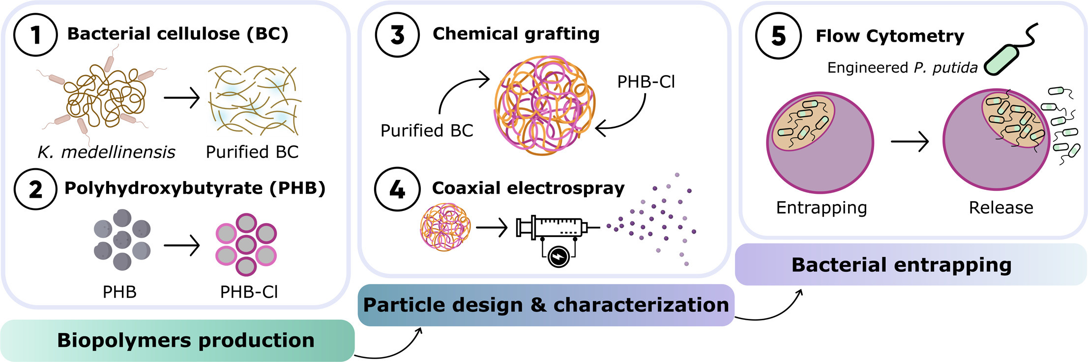
The design of biocarriers presents an effective approach for preserving bioactive elements and enabling controlled release in specific environments. This study introduces a novel biocarrier structure composed of two biodegradable, non-toxic, yet inherently incompatible bacterial biopolymers: bacterial cellulose (BC), a hydrophilic porous polymer known for its high water-holding capacity (up to 400 times its dry weight) and tensile strength, and polyhydroxybutyrate (PHB), a hydrophobic polymer characterized by its excellent barrier properties and UV stability. Using a coaxial electrospray technique, double-shelled hollow particles (DSHP) with a spherical architecture and an average diameter of 360 µm were produced. These particles consist of an outer PHB shell that serves as a protective barrier, and an inner BC-based layer designed to support microbial viability. To ensure structural integrity and enhance compatibility between the polymers, PHB chains were grafted onto BC, achieving a modification degree of 31%, prior to electrospraying. The resulting DSHP demonstrated an internal cavity capable of housing bacterial loads up to 108 CFU/mL, maintaining cell viability for at least 2 days and enabling controlled release profile. Additionally, the optimized electrospray conditions ensured high reproducibility and stability. This promising particle configuration offers potential applicability across various fields, from biomedicine to environmental applications.
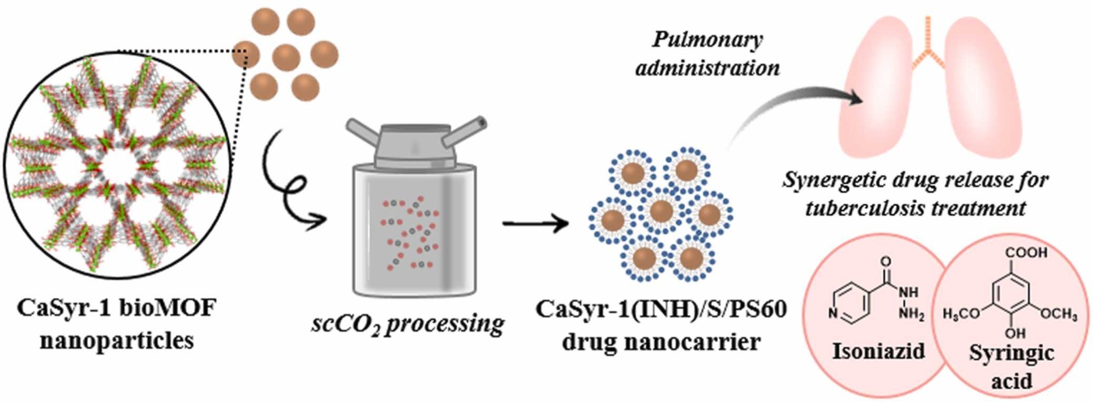
Supercritical CO2 assisted bioMOF drug encapsulation and functionalization for delivery with a synergetic therapeutic value

Despite the impressive characteristics of biological metal organic frameworks (bioMOFs) for their use as drug delivery systems (DDs), there are still some parameters related to their structural stability and processing routes that have decelerated their realistic application in this field. Both drawbacks are unraveled in this work for the microporous bioMOF CaSyr-1 by using supercritical CO2 (scCO2) to load the bioMOF with the anti-tubercular isoniazid (INH) drug, and functionalize its external surface with a hydrophobic protective layer of stearate (S). The hydrophobicized CaSyr-1(INH)/S vehicle is further coated with a neutral surfactant (PS60) to enhance the wettability of the system. In vitro tests, related to drug carrier biocompatibility and drug release in body simulated fluids, are performed to demonstrate potential prospective of the designed DDs in pharmacy. The synthetized product displayed total biocompatibility even at high concentrations, and the particle size and dissolution rate showed to be adequate for pulmonary administration.

BioMOF@cellulose Glycerogel Scaffold with Multifold Bioactivity: Perspective in Bone Tissue Repair

The development of new biomaterials for musculoskeletal tissue repair is currently an important branch in biomedicine research. The approach presented here is centered around the development of a prototypic synthetic glycerogel scaffold for bone regeneration, which simultaneously features therapeutic activity. The main novelty of this work lies in the combination of an open meso and macroporous nanocrystalline cellulose (NCC)-based glycerogel with a fully biocompatible microporous bioMOF system (CaSyr-1) composed of calcium ions and syringic acid. The bioMOF framework is further impregnated with a third bioactive component, i.e., ibuprofen (ibu), to generate a multifold bioactive system. The integrated CaSyr-1(ibu) serves as a reservoir for bioactive compounds delivery, while the NCC scaffold is the proposed matrix for cell ingrowth, proliferation and differentiation. The measured drug delivery profiles, studied in a phosphate-buffered saline solution at 310 K, indicate that the bioactive components are released concurrently with bioMOF dissolution after ca. 30 min following a pseudo-first-order kinetic model. Furthermore, according to the semi-empirical Korsmeyer-Peppas kinetic model, this release is governed by a case-II mechanism, suggesting that the molecular transport is influenced by the relaxation of the NCC matrix. Preliminary in vitro results denote that the initial high concentration of glycerol in the NCC scaffold can be toxic in direct contact with human osteoblasts (HObs). However, when the excess of glycerol is diluted in the system (after the second day of the experiment), the direct and indirect assays confirm full biocompatibility and suitability for HOb proliferation.

In vivo analysis of hybrid hydrogels containing dual growth factor combinations, and skeletal stem cells under mechanical stimulation for bone repair

Bone tissue engineering requires a combination of materials, cells, growth factors and mechanical cues to recapitulate bone formation. In this study we evaluated hybrid hydrogels for minimally invasive bone formation by combining biomaterials with skeletal stem cells and staged release of growth factors together with mechanotransduction. Hybrid hydrogels consisting of alginate and decellularized, demineralised bone extracellular matrix (ALG/ECM) were seeded with Stro-1+ human bone marrow stromal cells (HBMSCs). Dual combinations of growth factors within staged-release polylactic-co-glycolic acid (PLGA) microparticles were added to hydrogels to mimic, in part, the signalling events in bone regeneration: VEGF, TGF-β3, PTHrP (fast release), or BMP-2, vitamin D3 (slow release). Mechanotransduction was initiated using magnetic fields to remotely actuate superparamagnetic nanoparticles (MNP) targeted to TREK1 ion channels. Hybrid hydrogels were implanted subcutaneously within mice for 28 days, and evaluated for bone formation using micro-CT and histology. Control hydrogels lacking HBMSCs, growth factors, or MNP became mineralised, and neither growth factors, HBMSCs, nor mechanotransduction increased bone formation. However, structural differences in the newly-formed bone were influenced by growth factors. Slow release of BMP-2 induced thick bone trabeculae and PTHrP or VitD3 increased bone formation. However, fast-release of TGF-β3 and VEGF resulted in thin trabeculae. Mechanotransduction reversed the trabecular thinning and increased collagen deposition with PTHrP and VitD3. Our findings demonstrate the potential of hybrid ALG/ECM hydrogel–cell–growth factor constructs to repair bone in combination with mechanotransduction for fine-tuning bone structure. This approach may form a minimally invasive reparative strategy for bone tissue engineering applications.

Chemoselective reaction of methoxyaminomethyl BODIPYs with unprotected carbohydrates: a powerful tool for accessing BODIPY neoglycosides

The neoglycosylation of methoxyaminomethyl-appended BODIPYs with unprotected reducing mono-, di-, and trisaccharides produces, in a regio- and stereoselective manner, cyclic N-glycosyl-N-methoxy–BODIPY conjugates, as a relevant class of neoglycosides that display excellent photophysical characteristics in pure water, even at high dye concentrations. In addition, the cellular uptake of some of the neoglycosylated BODIPYs has been confirmed via fluorescence microscopy, and a BODIPY–acarbose conjugate showed comparable enzymatic inhibitory activity to acarbose for two different α-amylases: A. oryzae α-amylase (AOA) and human salivary α-amylase (HSA).

DOI
https://doi.org/10.1039/D4QO00886C

Europium–tannic acid nanocomplexes devised for bone regeneration under oxidative or inflammatory environments

Europium ions (Eu3+) are gaining attention in the field of regenerative medicine due to increasing evidence of their osteogenic properties. However, inflammatory and oxidative environments present in many bone diseases, such as osteoporosis or rheumatoid arthritis, are known to hinder this regenerative process. Herein, we describe a straightforward synthetic procedure to prepare Eu3+–tannic acid nanocomplexes (EuTA NCs) with modulable physicochemical characteristics, as well as antioxidant, anti-inflammatory, and osteogenic properties. EuTA NCs were rationally synthesized to present different contents of Eu3+ on their structure to evaluate the effect of the cation on the biological properties of the formulations. In all the cases, EuTA NCs were stable in distilled water at physiological pH, had a highly negative surface charge (ζ ≈ −25.4 mV), and controllable size (80 < Dh < 160 nm). In vitro antioxidant tests revealed that Eu3+ complexation did not significantly alter the total radical scavenging activity (RSA) of TA but enhanced its ability to scavenge H2O2 and ferrous ions, thus improving its overall antioxidant potential. At the cellular level, EuTA NCs reduced the instantaneous toxicity of high concentrations of free TA, resulting in better antioxidant (13.3% increase of RSA vs. TA) and anti-inflammatory responses (17.6% reduction of nitric oxide production vs. TA) on cultures of H2O2– and LPS-stimulated macrophages, respectively. Furthermore, the short-term treatment of osteoblasts with EuTA NCs was found to increase their alkaline phosphatase activity and their matrix mineralization capacity. Overall, this simple and tunable platform is a potential candidate to promote bone growth in complex environments by simultaneously targeting multiple pathophysiological mechanisms of disease.

DOI
https://doi.org/10.1039/D4TB00697F

Ti/Ta-based composite polysaccharide scaffolds for guided bone regeneration in total hip arthroplasty

Guided bone regeneration can play an important role in orthopedic applications. This work presents the synthesis and characterization of composite scaffolds based on polysaccharides loaded with microparticles of titanium or tantalum as novel materials proposed for composite systems with promising characteristics for guided bone regeneration. Ti/Ta composite scaffolds were synthesized using chitosan and gellan gum as organic substrates and crosslinked with oxidized dextran resulting in stable inorganic-organic composites. Physico-chemical characterization revealed a uniform distribution of metal nanoparticles within the scaffolds that showed a release of metals lower than 5 %. In vitro biological assays demonstrated that Ta composites exhibit a 2 times higher ALP activity than Ti and a higher capacity to support the full differentiation of human mesenchymal stem cells into osteoblasts. These results highlight their potential for bone regeneration applications.

1.
García-Robledo, H. et al. Ti/Ta-based composite polysaccharide scaffolds for guided bone regeneration in total hip arthroplasty. International Journal of Biological Macromolecules 271, 132573 (2024). Cite

Nanofibrous ε-Polycaprolactone Matrices Containing Nano-Hydroxyapatite and Humulus lupulus L. Extract: Physicochemical and Biological Characterization for Oral Applications

Oral bone defects occur as a result of trauma, cancer, infections, periodontal diseases, and caries. Autogenic and allogenic grafts are the gold standard used to treat and regenerate damaged or defective bone segments. However, these materials do not possess the antimicrobial properties necessary to inhibit the invasion of the numerous deleterious pathogens present in the oral microbiota. In the present study, poly(ε-caprolactone) (PCL), nano-hydroxyapatite (nHAp), and a commercial extract of Humulus lupulus L. (hops) were electrospun into polymeric matrices to assess their potential for drug delivery and bone regeneration. The fabricated matrices were analyzed using scanning electron microscopy (SEM), tensile analysis, thermogravimetric analysis (TGA), FTIR assay, and in vitro hydrolytic degradation. The antimicrobial properties were evaluated against the oral pathogens Streptococcus mutans, Porphyromonas gingivalis, and Aggregatibacter actinomycetemcomitans. The cytocompatibility was proved using the MTT assay. SEM analysis established the nanostructured matrices present in the three-dimensional interconnected network. The present research provides new information about the interaction of natural compounds with ceramic and polymeric biomaterials. The hop extract and other natural or synthetic medicinal agents can be effectively loaded into PCL fibers and have the potential to be used in oral applications.

Nanocarrier of α-Tocopheryl Succinate Based on a Copolymer Derivative of (4,7-dichloroquinolin-2-yl)methanol and Its Cytotoxicity against a Breast Cancer Cell Line

In order to improve the water solubility and, therefore, bioavailability and therapeutic activity of anticancer hydrophobic drug α-tocopherol succinate (α-TOS), in this work, copolymers were synthesized via free radicals from QMES (1-[4,7-dichloroquinolin-2-ylmethyl]-4-methacryloyloxyethyl succinate) and VP (N-vinyl-2-pirrolidone) using different molar ratios, and were used to nanoencapsulate and deliver α-TOS into cancer cells MCF-7. QMES monomer was chosen because the QMES pendant group in the polymer tends to hydrolyze to form free 4,7-dichloro-2-quinolinemethanol (QOH), which also, like α-TOS, exhibit anti-proliferative effects on cancerous cells. From the QMES-VP 30:70 (QMES-30) and 40:60 (QMES-40) copolymers obtained, it was possible to prepare aqueous suspensions of empty nanoparticles (NPs) loaded with α-TOS by nanoprecipitation. The diameter and encapsulation efficiency (%EE) of the QMES-30 NPs loaded with α-TOS were 128.6 nm and 52%; while for the QMES-40 NPs loaded with α-TOS, they were 148.8 nm and 65%. The results of the AlamarBlue assay at 72 h of treatment show that empty QMES-30 NPs (without α-TOS) produced a marked cytotoxic effect on MCF-7 breast cancer cells, corresponding to an IC50 value of 0.043 mg mL−1, and importantly, they did not exhibit cytotoxicity against healthy HUVEC cells. Furthermore, NP-QMES-40 loaded with α-TOS were cytotoxic with an IC50 value of 0.076 mg mL−1, demonstrating a progressive release of α-TOS; however, the latter nanoparticles were also cytotoxic to healthy cells in the range of the assayed concentrations. These results contribute to the search for a new polymeric nanocarrier of QOH, α-TOS or other hydrophobic drugs for the treatment of cancer or others diseases treatable with these drugs.

Facile, fast and green synthesis of a highly porous calcium-syringate bioMOF with intriguing triple bioactivity

A facile, fast and green strategy in ethanol is utilized to prepare a new bioMOF, namely CaSyr-1, with the particular characteristics of full biocompatibility given by using just calcium and syringic acid, the later being a phenolic natural product found in fruits and vegetables, permanent porosity with outstanding surface area >1000 m2g-1, and a micropore diameter of 1.4 nm close to mesopore values. Collectively, these data establish CaSyr-1 as one of the most porous bioMOFs reported to date, with high molecular adsorption capacity. The CaSyr-1 adsorptive behavior is revised here through the reversible adsorption of CO2 and the encapsulation of bioactive ingredients in the structure. Remarkably, CaSyr-1 enables the development of triple therapeutic entities, involving bioactive Ca2+, syringic acid and an impregnated drug.

1.
Rosado, A. et al. Facile, fast and green synthesis of a highly porous calcium-syringate bioMOF with intriguing triple bioactivity. Inorg. Chem. Front. 10, 2165–2173 (2023). Download Download
WP Twitter Auto Publish Powered By : XYZScripts.com